- Reception Numbers
- 0744-2790001
- 0744-2790002, 0744-2790003
Coarctation Of Aorta With Arch Hypoplasia

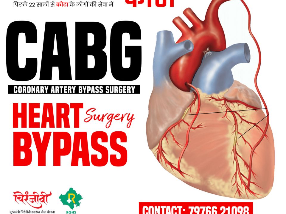
Coronary Artery Bypass Surgery | CABG | Heart Bypass Surgery in Kota
September 23, 2022- Best Cardiac Care Facility in Kota
- Best cardiac care Hospital In Kota
- Best cardiac care Medicine doctor in Kota
- Best Cardiac Care Services in Kota
- Best Cardiac Care Surgeon in Kota
- Best cardiac Hospital In Kota
- Best cardiology department in Kota
- Best Heart Hospital In Kota
- Best Heart Hospital iN Rajasthan
- Best Heart Surgeons in Kota
- Best Heart surgery in Kota
- Best Hospital for heart attack Treatment in Kota
- Best Hospital for heart surgery in Kota
- Best Hospital for heart Treatments in Kota
- Biggest Cardiology Facility in Kota
- Blue Baby
- Cardiac arrest surgery in Kota
- Cardiac care hospital in Kota
- Cardiac care services by RGHS
- Cardiac care services in Kota
- Cardiac Surgeon
- Cardiac Surgery by RGHS
- Coarctation Of Aorta
- Congenital Heart Disease
- Dr. Palkesh Agarwal Best Heart Surgeon in KOta
- Free Heart Bypass Surgery in Kota
- Heart Bypass Surgery Price in Rajasthan
- Heart Bypass Surgery Price in Sudha Hospital & Medical Research Centre
- Heart care services in Kota
- Heart Hospital Near Me
- Heart Hospitals in Kota
- heart operation by Mukhyamantri Chiranjivi Swasthya Bima Yojna in Rajasthan
- heart operation by Mukhyamantri Chiranjivi Swasthya Bima Yojna ki Kota
- Heart Operation by RGHS in Kota
- Heart Operation by RGHS in Rajasthan
- Heart problems in children
- Heart specialist in Kota
- Heart Surgeons in Kota
- Heart Surgery by RGHS
- Hole in Heart Surgery
- Infant Heart Surgery in Kota
- Most Advanced Heart Hospital in Kota
- Newborn Heart Surgery in Kota


What is Aortic Coarctation?
==========
Coarctation of the aorta is a narrowing of the aorta. The aorta is the main blood vessel carrying oxygen-rich blood from the left ventricle of the heart to all the organs of the body.
Coarctation occurs most often in a short piece of the aorta just beyond where the arteries to the head and arms take off.
This portion of the aorta is called the "juxtaductal" aorta.
The ductus arteriosus is a blood vessel that is normally present in a fetus. It has special tissue in its wall that causes it to close in the first hours or days of life. Coarctation may be caused by having extra ductal tissue.
In babies with coarctation, the aortic arch may also be small (hypoplastic). Coarctation may also occur with other cardiac defects, typically involving the left side of the heart. The defects usually seen with coarctation are bicuspid aortic valve and ventricular septal defect. Coarctation may also be seen as a part of more complex, single ventricle heart defects.
Coarctation of the aorta is common in some patients with genetic disorders, such as Turner's syndrome.
When a coarctation exists, the left ventricle must work harder to create a higher pressure than normal to force blood through the narrow part of the aorta to the lower part of the body.
If the narrowing is very severe, the ventricle may not be strong enough to do this extra work. This can lead to congestive heart failure or not enough blood flow to the organs of the body.
Diagnosis of Coarctation
====================
Coarctation of the aorta is present from birth. The age at which coarctation is found depends on the severity of the narrowing.
In about 25 percent of cases of isolated coarctation, the narrowing is severe enough to cause symptoms in the first days of life. When the ductus arteriosus closes, the left ventricle must suddenly pump against much higher resistance. This can lead to heart failure and shock. Because these newborns are well until the ductus arteriosus closes, symptoms appear quickly. They are often severe.
In patients who do not develop heart failure as newborns, coarctation may not be found until the child is several years old. In these older patients, coarctation is often first thought of because of a heart murmur or high blood pressure.
Coarctation is considered when the doctor is unable to feel pulses in a child’s legs. High blood pressure in the arms (but not the legs) may be noticed. A heart murmur is usually present. It may be loudest in the back. This is where the aorta is located.
The diagnosis of coarctation is confirmed with echocardiography. This can look at the anatomy of the aorta. It can also evaluate for other cardiac anomalies that may be present. Occasionally other tests, such as a cardiac MRI or CT scan, may be used to look at the coarctation.
Managing Coarctation
===================
In a critically ill newborn, the goals are to improve ventricular function and restore blood flow to the lower body. A continuous intravenous medication, prostaglandin (PGE-1), is used to open the ductus arteriosus. This allows blood flow to the body beyond the coarctation. Often other intravenous (IV) medicines may also be needed to help the heart. Many babies need to be placed on a ventilator before surgery.
If the baby has symptoms of a coarctation, surgery is done on an urgent basis.
There are a few surgical techniques to repair coarctation. The most common repair involves resection (removal) of the narrowed area with anastomosis (reconnection) of the two ends to each other. Sometimes the resection (removal) must be extended towards the arch if there is a longer piece of narrowing. In another method, the narrowing may be opened with a patch. Or a portion of an artery may be used as a flap to expand the area (called a subclavian flap aortoplasty).
Because older children may have minimal symptoms, coarctation repair is typically planned electively. Surgical repair is usually done with resection of the narrowed piece and end-to-end reconnection.
In older children, transcatheter therapy is the first-line therapy. It offers the ability to use a balloon or stent to dilate (make the area bigger) the area of narrowing without needing surgery. In children who are still growing, the placement of a stent means that an additional catheterization procedure is likely to be needed in the future.
Why to choose Sudha Hospital & Medical Research Centre for Heart Treatments?
Answer: Sudha Hospital is equipped with state of the art technology, We do have CATH Lab, ICU and all other necessary arrangements for emergency situations. All tests available under one roof. We have highly experience cardiac physicians and cardiac surgeon who are offering round the clock services in Kota. We are famous for new born heart surgeries. With the grace of god the success rate of Paediatric Heart Surgery is extremely high at Sudha Hospital & Medical Research Centre.
website: https://sudhahospitalkota.com/


